Cardiac Catheterization

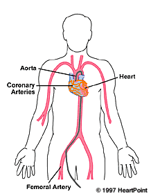
Cardiac catheterization goes by a variety of names: "Heart cath", "Cardiac cath", "Coronary angiogram" or "angiography". In any case, the procedure performed uses "catheters", which are long, thin and flexible tubes that are introduced from the arm or leg and passed through the blood vessels to the heart. It can be useful in diagnosing almost all types of heart disease. Congenital heart disease, diseases of the heart valves and heart muscle are evaluated by the measurement of pressures, blood flow directions and quantities, and pictures of heart muscle function. The most common problem with hearts in developed countries are those of the coronary arteries which supply the heart muscle with blood. Cardiac catheterization which includes evaluation of these structures involves the injection of dye into the vessels.
The coronary arteries arise from the aorta, and travel along the outside of the heart, bringing blood containing oxygen and necessary nutrients to the heart muscle. These are the structures seen during coronary arteriography. For more details, see "Coronary Artery Disease".
The following section is designed to help people understand various aspects of the procedure. Be sure to ask your physician regarding the specifics of your own particular case.
| Does everyone with heart disease need a heart cath? | Absolutely not. In fact, cardiac
catheterization may add little to the diagnosis and management of many cardiac conditions.
It is expensive, somewhat time-consuming, and does have some risks (see below). However, it is essential for the diagnosis and treatment of many of the most serious forms of heart disease, including many cases coronary artery disease, the most common form of lethal heart problems. Even then, there may be alternative modes of diagnosis. It is beyond the scope of virtually any document to discuss all of the possible variations that are possible, since every case in individualized. Suffice it to say here that the precision of the diagnostic information obtained may well be worth the time, risk and expense in many cases. |
| What is done before the procedure? | Many catheterizations these days are scheduled as an
"outpatient procedure", and the patient is not formally admitted to the
hospital. In this case, you will generally come to the hospital or other location where
the procedure is to be done on the morning of the procedure. It is generally recommended that no food or drink should be taken for 6-8 hours prior to the procedure to minimize nausea or vomiting (most physicians allow a sip of water to take medications). If you are having an outpatient procedure, be sure you have brought someone who can drive you home later that day. You should pack a small bag in case you need to stay at the hospital after the procedure. Generally, routine blood tests and an electrocardiogram will be done either before you come to the hospital or shortly after you arrive. A small intravenous line will generally be started in the hand or arm. The hair will be shaved from the site where the catheter will be inserted. You will generally be allowed to wear glasses, hearing aids and dentures. |
| Where is the procedure done?
|
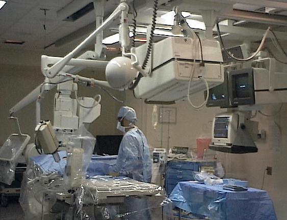
Most hospitals have special rooms referred to as
"cath labs". (there are even mobile cath labs which are self-contained in
semi-trucks traveling from location to location). They contain a table for the patient,
x-ray tubes, and equipment to monitor the heartbeat and blood pressure. In addition to the
physician performing the test, there is generally at least one technician or nurse who
assists, and one who monitors various parameters who is outside the room itself. |
| How long does the procedure take? | The actual catheterization procedure takes about 15-30 minutes. This is the time it takes the physician to perform the procedure. It takes time before this to get to the room, have the skin prepared and draped, etc. |
| What preparations occur in the cath lab when I first get in there? | You will be placed on a long slender bed (this is more like a table and is designed for getting good pictures of the arteries, not comfort necessarily!). The area where the catheter will be inserted will be scrubbed, and the area draped. Electrocardiographic leads will be placed so that the heart rhythm can be monitored, and a blood pressure cuff may be placed as well. Oxygen is sometimes administered. A device to monitor the concentration of oxygen may be gently clipped to the finger or ear. A sedative may be administered (see below). |
| Will I be awake? | Catheterization is generally considered a "minor
procedure" from the surgical standpoint, and sedation and local anesthesia are used.
Some people "don't want to know anything" and would like to be "knocked out
totally" just like undergoing anesthesia for a major surgery. The risk of general
anesthesia is not felt to be justified for this procedure. Most physicians give some sedation, but the amount is quite variable. Some use a relatively small amount of oral sedatives (relaxing medications), while others give fairly substantial amounts of these or similar agents intravenously. In the first situation, the patient will generally be awake and aware during the procedure. In the second case, the patient may be very sedated, be able to breath on their own, but have little memory of the procedure. You should let the physician performing the test know your preference. |
| Does it hurt? | In most cases, only mild discomfort is involved. An
occasional patient will have quite a bit of pain (and it seems they are the ones who tell
the most people!). Although it may sound as though it will be pretty painful, most people
tolerate the procedure very well. The site where the catheters are to be placed with be numbed with a local anesthetic agent, similar to ones used by dentists. There are fewer pain fibers in the leg and groin than in the mouth, so the discomfort is generally less than one experiences in the dentist's chair. Once the catheters are in place, there is generally very little discomfort. People undergoing the procedure do not feel the catheters move through the blood vessels on their way to the heart. You may feel an occasional heart "skip" during the procedure. In many procedures, a fairly substantial amount of dye is injected to visualize the contraction of the heart muscle. The dye which is injected is often perceived as warm, but is not often referred to as painful. An occasional patient will experience discomfort with injections of dye into the heart arteries, but this is quite uncommon. |
| How do they get the catheters to the right spot? | It's done with the aid of x-rays which can follow the catheter's course, and it's generally a lot easier than it may seem at first. The arteries and veins all connect pretty directly to the heart. It's a matter of getting on the main highway . . . the rest of the journey is usually pretty straightforward (some people with blocked arteries in their legs or whose vessels are less straight do present a bit of a challenge). |
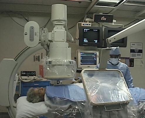
| Can I watch? | Most catheterization laboratories are set up so that the
person undergoing the procedure can watch parts of the procedure on the x-ray screen if
they so desire. The x-ray tube that makes the pictures sometimes blocks the patient's
view, however. If you don't want to watch, you may close your eyes! |
| What is the dye made of? | The "dye" really isn't a dye, and doesn't
"stain" anything. The dye looks clear to the naked eye. To the x-ray machine,
the liquid, which contains iodine, appears white. The dye is filtered by the kidneys and excreted in the urine. |
| What do the pictures look like? Why do they take so many? | Since the heart is always moving, the pictures made in the cath lab are generally moving pictures. They are recorded on some combination of videotape, 35 mm film, or stored as digital computerized images. There are many branches of the coronary arteries, and their images cross over each other in the two dimensional views that are made. Therefore, different angles must be taken to visualize all of the segments. |
| When will I know the results? | Often, a precise diagnosis is available immediately. In other cases, other calculations may need to be done on some of the data obtained, films developed for further review, or consultations with other physicians requested. The physician performing the procedure will generally give at least some preliminary indications to you and your family shortly after the procedure is completed. |
| What if they find blockages? Will they balloon them out right then? | It depends on a number of factors: Some catheterization laboratories do not allow both procedures to be done at
once, since this increases the unpredictability of their schedules. |
| Are there risks? | There are risks associated with catheterization. They are
fairly infrequent. The risk of the complications is generally greater when the patient is
sicker This is often the same time as when the procedure is most needed. The risk of major complications is approximately 1 in 1000. The risk of death is 1 in 5000. Several specific risks are discussed below. Bleeding. Whenever the skin is entered, there is a risk of bleeding. Mild blood loss occurs with all procedures. More severe bleeding may occur on occasion, generally at the site where the catheters are inserted. Most often, this can be controlled without much fuss. Occasionally, there is enough blood loss that transfusion of blood is required. Surgery is rarely needed to control excessive bleeding. Infection. There is also always the risk of infection with any surgical procedure. The risk of infection with this procedure is extremely low. Injury to a nerve. Arteries and veins generally travel along with nerves, and therefore irritation, inflammation, or injury can occur to these structures. When it does occur in procedures done from the groin, it generally leads to numbness along the leg to the knee. This is usually temporary. Stroke. A stroke occurs when there is a loss of blood flow to part of the brain. This may occur in association with a heart catheterization if a small amount of "plaque" is dislodged from the inside of the blood vessels through which the catheters are being passed, if a clot is formed on a catheter and is dislodged, or if clot already present in the heart is dislodged. Bubbles of air accidentally released into the blood stream may lodge in a blood vessel and interrupt the flow of blood. Careful attention to avoid allowing these events to occur, but not all such events can be prevented. Heart attack. Heart attacks can also occur. Heart caths are surprisingly safe in patients with significant blockages of the heart arteries, but heart attacks do occur during the procedure. These are mostly the real "heart attacks waiting to happen", and one could argue there's no better place to have a heart attack than in the cath lab where it can be promptly tended to. Death. Deaths occur in the cath lab. Most deaths that do occur happen in patients who are extremely ill before they get to the lab, and are being brought there in desperate circumstances. While very rare, deaths do occur in patients who have no heart disease but have a complication of the procedure. Injuries to blood vessels. Injuries to the blood vessels where the catheters are inserted certainly can occur. Some complications, such as a "pseudoaneurysm" are diagnosed and often treated with ultrasound techniques. Surgery is sometimes required to remove a clot from the leg, repair a torn blood vessel or other problems. Kidney damage. The contrast material used in these procedures can be tough on the kidneys. People with previous kidney problems, and those with diabetes are particularly at risk, although problems occasionally occur in people without either of these conditions. The kidneys may stop filtering fluids or excreting toxic substances in the urine. Kidney failure may result, and dialysis (either temporary or permanent) may occur. There are precautions that may be taken to minimize the chance of kidney injury, particularly in high-risk patients. Radiation. The x-ray exposure from this procedure is not expected to be harmful. |
| This is scary. Should I really have the procedure? | Indeed, after hearing of these risks, one can feel scared
a little or a lot. There are two things to remember: The
frequency of complications is very low, as noted above. |
| Are there alternatives? | In discussions of risks and benefits, one also has to
consider the alternatives to catheterization. Depending on the circumstances,
"non-invasive" studies such as echocardiograms, treadmill stress tests, nuclear
cardiology studies, and other techniques may provide important information about the
heart. Each of these have a role in certain circumstances, a subject which is well beyond
the scope of this discussion. Undoubtedly, your physician has considered these
alternatives in your particular situation, but you will need to ask him/her specifically
why these alternatives are not felt to be more appropriate in your case. Remember, you are ultimately the one who decides on which test, if any, is done to you! You should not feel uncomfortable at all about asking. |
| What other procedures are done in the cath lab? | Related procedures, which are nevertheless different,
include Carotid angiography at which time angiograms are made of
the arteries to the brain. The heart is not entered, and pictures of the heart arteries
are not made. |
| How do they stop the site from bleeding? | Usually, only pressure and bed rest are all that are
required. This is because most procedures are done with a "percutaneous needle"
technique. This just means that a needle is introduced through the skin without direct
visualization of the artery. A relatively small hole is made, and when the catheters are
removed, the blood vessels which have muscle layers will simply contract and close the
wound. Pressure is held on the site to help with closure and the formation of a clot. Some centers now use a variety of techniques to stop bleeding at the site by suturing the wound close with special tools that accomplish this without direct visualization of the hole they are closing. Fibrin "plugs" can also be placed. Both of these techniques may often, but not always, shorten the time of bed rest which is required. Since the technology adds a substantial amount to the costs, and the benefits are not substantial in the eyes of many, they are not widely used at this time. A sandbag may be placed over the site. A bulky pressure dressing, or a simple bandage, may be applied to the site. If a "cutdown" procedure has been performed, the vessels are closed surgically and less bed rest is required afterwards. |
| When can I eat and drink? | Important questions!! You may generally eat and drink shortly after the procedure. |
| What about after I leave the hospital? | You will need to limit your activity a bit for the 24
hours or so after the procedure. You can certainly move about and do your activities of
daily living. However, you should avoid heavy lifting or straining. Your doctor should
tell you when you may resume your normal activities. You generally can take off the dressing the day following the procedure. Showering and bathing generally can be resumed at this time as well. You will more than likely have a bruise at the site of the catheterization. It may be small, or rather spectacular -- even a little bit of blood in the deeper tissues can travel a long way, and may surface on the skin at the knee! The size of the bruise alone is important only to your appearance. It will slowly resolve over time. It may be a bit painful, as the white blood cells work on digesting the blood making up the bruise. If there is a drop or two of blood on the dressing, this is generally not a concern. However, if more than this occurs, put pressure on the site with your hand, and seek medical attention. Call your doctor if you have marked pain at the site. If there are signs of infection (this would be most unusual), let your doctor know as well. |
| I had a reaction to iodine or shellfish before. What should I do? | You should let your physician know. Iodine is used as the basis for the contrast materials ("dye") used in these procedures. These substances have been refined greatly over the years, and they now are less toxic to the kidneys, cause less nausea, and fewer problems with irregular heartbeats. Allergies to iodine are not uncommon, but seem to be less frequent with the more modern contrast agents. It is important to let your physician know if you are allergic to iodine or if you have had previous reactions to contrast agents. Pretreatment with certain medications can markedly reduce the frequency and severity of any possible reactions. Let your physician know also if you have reactions or are allergic to shellfish or seafood,. These foods have large amounts of iodine, and pretreatment may be indicated to avoid possible problems. |
©COPY;1997 HeartPoint Updated November 1998
Please see these related areas on HeartPoint:
| Commentary |
Food You Will Love | HeartPoint
Gallery | In The News | Health Tips | What's New
|
| Information Center | Home
|
This site presents material for your information, education and entertainment. We can assume no liability for inaccuracies, errors, or omissions. Above all, material on this site should not take the place of the care you receive from a personal physician. It is simply designed to help in the understanding of the heart and heart disease, and not as a diagnostic or therapeutic aid. You should seek prompt medical care for any specific health issues. Please feel free to browse the site and download material for personal and non-commercial use. You may not however distribute, modify, transmit or reuse any of these materials for public or commercial use. You should assume that all contents of the site are copyrighted. ©COPY;1997 HeartPoint