HeartPoint animation: this will take approximately 1 minute to load.

Valvular Heart Disease
HeartPoint animation: this will take approximately 1 minute to
load.

It is easily understood that the muscle that we call the heart must continue to pump with adequate force to pump the blood that the body needs. "Valves" however are extremely important to the heart's efficiency. These delicate structures allow for the efficient flow of blood progressively forward through the heart's chambers, maximizing the efficiency of the heart muscle's work.
To review the flow of blood through the heart, you can check out "The Heart" animation. link
In the animation above, the Tricuspid Valve (between the right atrium and right ventricle) and the Pulmonic Valve (between the right ventricle and the pulmonary artery) are illustrated to be working normally. After the right ventricle contracts, pressure is low in the chamber. The Tricuspid Valve, which had been closed from the pressure generated from the ventricle's contraction, now opens as the pressure of the blood from the right atrium has built up while the Tricuspid Valve was closed. The right ventricle will again contract, closing the Tricuspid Valve again, and pushing open the Pulmonic Valve. Once the right ventricle completes its contraction, the pressure in the pulmonary artery will be higher than in the right ventricle, and the Valve will close.
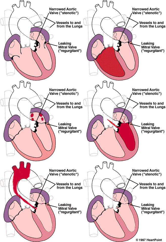
The valves on the left side of the heart, the Aortic Valve and the Mitral Valve however, are not working properly. Blood returns from the lungs and empties into the left atrium. In this illustration, the Mitral Valve opens properly when the left ventricle is finished contracting, and allows blood to flow into the left ventricle easily. When the left ventricle contracts however, blood is shown to flow back into the left atrium through the Mitral Valve. This backward flow of blood is called "regurgitation" or "insufficiency". Since it occurs through the Mitral Valve, it is termed "Mitral Regurgitation" or "Mitral Insufficiency".
The other valve on the left side of the heart, the Aortic Valve, is also illustrated to have the other main problem associated with heart valves. In this case, the valve is thickened and perhaps calcified. Instead of being easy to open as the left ventricle contracts, the left ventricle must push very hard to open the valve and allow blood to flow out to the rest of the body. This difficulty in opening the valve is called "stenosis", and since it involves the Aortic Valve, is called "Aortic Stenosis".
To learn more about the heart valves in health and disease, select "Tell Me More".
©COPY 1997 HeartPoint Updated May 1998
| Commentary |
Food You Will Love | HeartPoint
Gallery | In The News | Health Tips | What's New
| Information Center | Home
|
This site presents material for your information, education and entertainment. We can assume no liability for inaccuracies, errors, or omissions. Above all, material on this site should not take the place of the care you receive from a personal physician. It is simply designed to help in the understanding of the heart and heart disease, and not as a diagnostic or therapeutic aid. You should seek prompt medical care for any specific health issues. Please feel free to browse the site and download material for personal and non-commercial use. You may not however distribute, modify, transmit or reuse any of these materials for public or commercial use. You should assume that all contents of the site are copyrighted. ©COPY;1997 HeartPoint